April 24, 2007
A closer look
Share this story
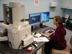
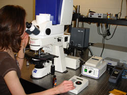
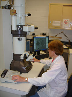
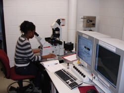
Virginia Commonwealth University researchers from a broad spectrum of disciplines armed with the latest microscope technology are revealing secrets of sub-cellular worlds in the Microscopy Facility of the Department of Anatomy and Neurobiology.
Some of these researchers are investigating the cellular responses involved in brain injury and how the brain works. Some are looking at the cellular players involved with inflammatory responses and wound repair. Yet others are examining how tumors grow and proliferate.
Their findings could have potential implications in the treatment and prevention of disease.
VCU researchers including neurobiologists, cell biologists, cancer researchers, biochemists, bioengineers, dental researchers, geneticists, physiologists and microbiologists, have access to nine different microscopy systems with a variety of advanced functions and capabilities. The facility, housed in the VCU School of Medicine, offers the equipment and expertise to examine sub-cellular details at high-resolution, enabling researchers to take their work to the next level.
Technology has dramatically advanced microscopes and enhanced what researchers can see and use for both qualitative and quantitative analysis. Scientists now can examine an organism’s life on the most intimate level and investigate the molecular dynamics and interactions within living cells.
The facility offers fluorescence, confocal and multi-photon laser scanning, transmission electron and scanning electron microscopes, each with its own dedicated environment and sample preparation stations. Advanced software applications are also available for quantitative image analysis. Expert staff is available for consultation, instruction, assistance, and scientific collaboration.
“This microscopy resource is amongst the most comprehensive in the Commonwealth of Virginia. It’s an excellent facility that has attracted top-rate faculty to VCU,” said Scott Henderson, Ph.D., the director of the Microscopy Facility. “We are always looking at ways to improve our facility and future plans for development are based upon trends in research.”
“As an example, we have just upgraded our two-photon system to include simultaneous physiological recording with imaging capabilities and a new total internal reflection fluorescence imaging system is currently being installed in the facility,” he said.
The Microscopy Facility has been made possible through grants from the National Institute of Neurological Disorders, the National Center for Research Resources and support from VCU and the VCU School of Medicine, and the Center for Disease Control.
For more information, visit http://www.vcu.edu/anatomy/microscopy/index.shtml.
Subscribe to VCU News
Subscribe to VCU News at newsletter.vcu.edu and receive a selection of stories, videos, photos, news clips and event listings in your inbox.