Small wonders: The microscopic images currently on display at the Tompkins-McCaw Library prove that the beauty of science is in the details

Share this story

A black widow spider’s silk-spinning organ. An ovarian tumor. Muscle fibers in the inner ear. Vagus neurons in a zebrafish’s brain. A traumatic brain injury in a mouse.

These are just a few examples of the microscopic images on display as part of the “Through the Looking Glass” art exhibit at the Tompkins-McCaw Library for the Health Sciences at Virginia Commonwealth University.

The exhibit, which is open to the public and will run through Dec. 31, showcases colorful and striking images created by VCU students, faculty and staff – all through the lens of a microscope.

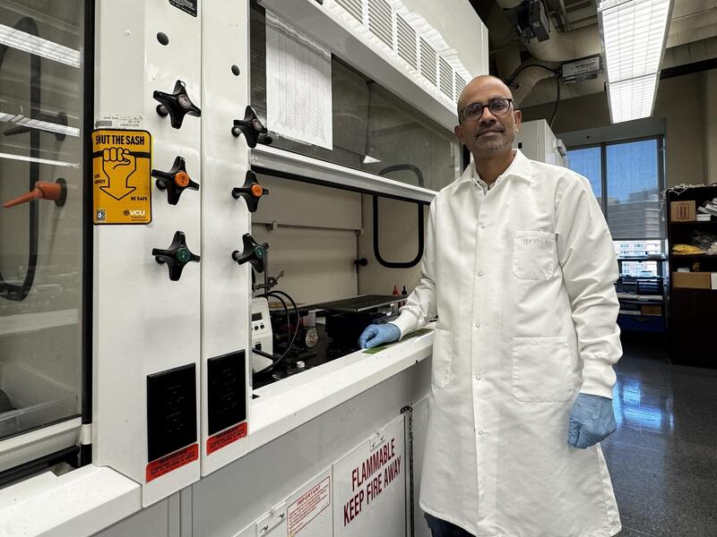
Last spring, a panel of judges from VCU’s biomedical engineering and clinical laboratory sciences, led by Scott Henderson, Ph.D., director of VCU’s Microscopy Facility, which is part of the Department of Anatomy and Neurobiology in the School of Medicine, chose the exhibit’s 24 images from 40 submissions, and selected three images as prizewinners on the basis of aesthetic appeal, technical skill and scientific significance.

“Exhibited images span disciplines from biomedical engineering to forensic science to pathology and range from scaffolding from electrospun fibers to a black widow spider spinnerets to a time-lapse image of vagus neurons,” said Teresa L. Knott, director of Tompkins-McCaw Library. “The images selected for the exhibit are dynamic and fascinating to me.”

The following is a selection of images from “Through the Looking Glass,” along with brief descriptions of the subject matter depicted and its scientific relevance.

<p style="text-align: left;"><span style="font-family: sans-serif; font-size: large;"><strong>“My Little Jellyfish”</strong></span><br>
<em>Kelly Hotchkiss, student, Department of Biomedical Engineering, School of Engineering</em></p>
<p style="text-align: left;"><span style="font-family: calibri;">Hotchkiss’ image shows human mesenchymal stem cells that present a spread pattern of attachment after being prepared on a glass surface.</span>

“My Little Jellyfish”
Kelly Hotchkiss, student, Department of Biomedical Engineering, School of Engineering

Hotchkiss’ image shows human mesenchymal stem cells that present a spread pattern of attachment after being prepared on a glass surface.

<p style="text-align: left;"><span style="font-family: sans-serif; font-size: large;"><strong>“A Serous Borderline Ovarian Tumor With Micropapillary Features”<br /></strong></span><em>Ema Dragoescu, M.D., assistant professor, </em><em><a href="http://www.pathology.vcu.edu/index.html" target="_blank"><span style="color: blue; font-family: calibri;"><span style="text-decoration: underline;">Department of Pathology</span></span></a><span style="font-family: calibri;">, School of Medicine</span></em></p>
<p style="text-align: left;"><span style="font-family: calibri;">This image is of a hematoxylin eosin-stained section of an ovarian tumor at 200-times magnification. The cells surrounding the central core form thin, filiform micropapillae – an arrangement known as the “Medusa head” – that indicate aggressive behavior in the tumor.</span>

“A Serous Borderline Ovarian Tumor With Micropapillary Features”
Ema Dragoescu, M.D., assistant professor, Department of Pathology, School of Medicine

This image is of a hematoxylin eosin-stained section of an ovarian tumor at 200-times magnification. The cells surrounding the central core form thin, filiform micropapillae – an arrangement known as the “Medusa head” – that indicate aggressive behavior in the tumor.

<p style="text-align: left;"><span style="font-family: sans-serif; font-size: large;"><strong>“Actin Reorganization in Transgenic Mouse Lung Cells”<br /></strong></span><em>Shilpa Singh, student, </em><em><a href="http://www.biochemistry.vcu.edu/" target="_blank"><span style="color: blue; font-family: calibri;"><span style="text-decoration: underline;">Department of Biochemistry and Molecular Biology</span></span></a><span style="font-family: calibri;">, School of Medicine</span></em></p>
<p style="text-align: left;"><span style="font-family: calibri;">These transgenic mice lung cells were stained using phalloidin and mounted using DAPI – a fluorescent stain – at 40-times magnification.</span>

“Actin Reorganization in Transgenic Mouse Lung Cells”
Shilpa Singh, student, Department of Biochemistry and Molecular Biology, School of Medicine

These transgenic mice lung cells were stained using phalloidin and mounted using DAPI – a fluorescent stain – at 40-times magnification.

<p style="text-align: left;"><span style="font-family: sans-serif; font-size: large;"><strong>“A Gathering of Osteoblasts”<br /></strong></span><em>Gireesh Reddy, student, Department of Biomedical Engineering, School of Engineering</em></p>
<p style="text-align: left;"><span style="font-family: calibri;">This image aims to provide insight into the distribution of integrin alpha V (stained in blue) in the cell.</span>

“A Gathering of Osteoblasts”
Gireesh Reddy, student, Department of Biomedical Engineering, School of Engineering

This image aims to provide insight into the distribution of integrin alpha V (stained in blue) in the cell.

<p style="text-align: left;"><span style="font-family: sans-serif; font-size: large;"><strong>“Visualizing Vision”<br /></strong></span><em>Paul Vorster, Ph.D. student, </em><em><a href="http://www.vcu.edu/lifesci/phd/" target="_blank"><span style="color: blue; font-family: calibri;"><span style="text-decoration: underline;">Integrative Life Sciences</span></span></a><span style="font-family: calibri;">, </span><a href="http://www.vcu.edu/lifesci/" target="_blank"><span style="color: blue; font-family: calibri;"><span style="text-decoration: underline;">VCU Life Sciences</span></span></a></em></p>
<p style="text-align: left;"><span style="font-family: calibri;">“Visualizing Vision” shows an array of retina cells surrounding the lens during eye development in a zebrafish embryo. The image was taken at the embryonic developmental stage relevant to retina and lens development in zebrafish at 24 hours post-fertilization.</span>

“Visualizing Vision”
Paul Vorster, Ph.D. student, Integrative Life Sciences, VCU Life Sciences

“Visualizing Vision” shows an array of retina cells surrounding the lens during eye development in a zebrafish embryo. The image was taken at the embryonic developmental stage relevant to retina and lens development in zebrafish at 24 hours post-fertilization.

<p style="text-align: left;"><span style="font-family: sans-serif; font-size: large;"><strong>“Electrospun Fibers”<br /></strong></span><em>Ryan Clohessy, student, Department of Biomedical Engineering, School of Engineering</em></p>
<p style="text-align: left;"><span style="font-family: calibri;">This image shows electrospun fibers that were created in an effort to develop a scaffold suitable for tissue engineering. The image was one of three selected as prizewinners by the judges.</span></p>
<p style="text-align: left;"><span style="font-family: calibri;">“I think combining science and artistic creativity has led to some amazing images and hope the beauty that can be found in science continues to be recognized and appreciated,” Clohessy said, after his image was selected as a prizewinner.</span>

“Electrospun Fibers”
Ryan Clohessy, student, Department of Biomedical Engineering, School of Engineering

This image shows electrospun fibers that were created in an effort to develop a scaffold suitable for tissue engineering. The image was one of three selected as prizewinners by the judges.

“I think combining science and artistic creativity has led to some amazing images and hope the beauty that can be found in science continues to be recognized and appreciated,” Clohessy said, after his image was selected as a prizewinner.

<p style="text-align: left;"><span style="font-family: sans-serif; font-size: large;"><strong>“Ear”</strong></span><br /><em><a href="http://biology.vcu.edu/people/robert-tombes" target="_blank"><span style="color: blue; font-family: calibri;"><span style="text-decoration: underline;">Robert Tombes</span></span></a><span style="font-family: calibri;">, Ph.D., professor, </span><a href="http://biology.vcu.edu/" target="_blank"><span style="color: blue; font-family: calibri;"><span style="text-decoration: underline;">Department of Biology</span></span></a><span style="font-family: calibri;">, </span><a href="http://www.has.vcu.edu/" target="_blank"><span style="color: blue; font-family: calibri;"><span style="text-decoration: underline;">College of Humanities and Sciences</span></span></a></em></p>
<p style="text-align: left;"><span style="font-family: calibri;">“Ear” shows muscle fibers (in green) and tubulin (in red), portraying the “axonal innervation of the ear into sensory structures.” According to the exhibit’s display, the image “suggests novel ways to regrow auditory sensors within the inner ear.”</span>

“Ear”
Robert Tombes, Ph.D., professor, Department of Biology, College of Humanities and Sciences

“Ear” shows muscle fibers (in green) and tubulin (in red), portraying the “axonal innervation of the ear into sensory structures.” According to the exhibit’s display, the image “suggests novel ways to regrow auditory sensors within the inner ear.”

<p style="text-align: left;"><span style="font-family: sans-serif; font-size: large;"><strong>“The Universal Thinker”<br /></strong></span><em>Ness Sufrin, student, </em><em><a href="http://www.has.vcu.edu/forensics/" target="_blank"><span style="color: blue; font-family: calibri;"><span style="text-decoration: underline;">Department of Forensic Science</span></span></a><span style="font-family: calibri;">, College of Humanities and Sciences</span></em></p>
<p style="text-align: left;"><span style="font-family: calibri;">This image displays a mixture of salt crystals that have been melted and are in the midst of recrystallizing.</span>

“The Universal Thinker”
Ness Sufrin, student, Department of Forensic Science, College of Humanities and Sciences

This image displays a mixture of salt crystals that have been melted and are in the midst of recrystallizing.

<p style="text-align: left;"><span style="font-family: sans-serif; font-size: large;"><strong>“Nanoscale Corals”<br /></strong></span><em>Ahmed A. Farghaly, student, </em><em><a href="https://chemistry.vcu.edu/" target="_blank"><span style="color: blue; font-family: calibri;"><span style="text-decoration: underline;">Department of Chemistry</span></span></a><span style="font-family: calibri;">, College of Humanities and Sciences</span></em></p>
<p style="text-align: left;"><span style="font-family: calibri;">“Nanoscale Corals” shows coral-like gold nanostructures that were prepared from silica-gold nanocomposite materials using a newly developed electroassisted codeposition approach. The high surface areas and nanosized features of the structures are ideal materials for many technological applications, including chemical sensing, biosensing and catalysis.</span>

“Nanoscale Corals”
Ahmed A. Farghaly, student, Department of Chemistry, College of Humanities and Sciences

“Nanoscale Corals” shows coral-like gold nanostructures that were prepared from silica-gold nanocomposite materials using a newly developed electroassisted codeposition approach. The high surface areas and nanosized features of the structures are ideal materials for many technological applications, including chemical sensing, biosensing and catalysis.

<p style="text-align: left;"><span style="font-family: sans-serif; font-size: large;"><strong>“Smiling Cells”<br /></strong></span><em>Tricia Hardt Smith, faculty instructor, Department of Biology, College of Humanities and Sciences</em></p>
<p style="text-align: left;"><span style="font-family: calibri;">Smith’s entry is the first picture to confirm the presence of Cannabinoid Receptor Interacting Protein 1a in gastrointestinal cells. The protein is stained in green while the blue represents cell nuclei. CRIP1a has previously been characterized in brain, eye and transfected cell culture systems.</span></p>
<p style="text-align: left;"><span style="font-family: calibri;">“CRIP1a has been shown to bind to the C-terminus of cannabinoid (CB1) receptors and inhibit constitutive CB1 receptor activity,” Smith said. “Cannabinoid receptors are found in the gut, and it is possible that CRIP1a is mediating receptor activity in the gut as well the brain.”</span>

“Smiling Cells”
Tricia Hardt Smith, faculty instructor, Department of Biology, College of Humanities and Sciences

Smith’s entry is the first picture to confirm the presence of Cannabinoid Receptor Interacting Protein 1a in gastrointestinal cells. The protein is stained in green while the blue represents cell nuclei. CRIP1a has previously been characterized in brain, eye and transfected cell culture systems.

“CRIP1a has been shown to bind to the C-terminus of cannabinoid (CB1) receptors and inhibit constitutive CB1 receptor activity,” Smith said. “Cannabinoid receptors are found in the gut, and it is possible that CRIP1a is mediating receptor activity in the gut as well the brain.”

<p style="text-align: left;"><span style="font-family: sans-serif; font-size: large;"><strong>“Epithelial”<br /></strong></span><em>Roshni Malik, student, Department of Biomedical Engineering, School of Engineering</em></p>
<p style="text-align: left;"><span style="font-family: calibri;">Many cells are depicted in Malik’s image, demonstrating the presence of fibronectin in green. The darker regions are cell nuclei. This image is used in breast cancer research to explore the role of fibronectin in epithelial to mesenchymal transition.</span>

“Epithelial”
Roshni Malik, student, Department of Biomedical Engineering, School of Engineering

Many cells are depicted in Malik’s image, demonstrating the presence of fibronectin in green. The darker regions are cell nuclei. This image is used in breast cancer research to explore the role of fibronectin in epithelial to mesenchymal transition.

<p style="text-align: left;"><span style="font-family: sans-serif; font-size: large;"><strong>“The Abdomen of the Southern Black Widow”<br /></strong></span><em><span style="font-family: calibri;">Ryan Clohessy, student, </span><a href="http://biomedical.egr.vcu.edu/" target="_blank"><span style="color: blue; font-family: calibri;"><span style="text-decoration: underline;">Department of Biomedical Engineering</span></span></a><span style="font-family: calibri;">, </span><a href="http://www.egr.vcu.edu/" target="_blank"><span style="color: blue; font-family: calibri;"><span style="text-decoration: underline;">School of Engineering</span></span></a></em></p>
<p style="text-align: left;"><span style="font-family: calibri;">This image is a dorsal view of latrodectus mactans, or the southern black widow spider. The spider’s spinnerets – its silk-spinning organ – can be seen at the base of the abdomen.</span>

“The Abdomen of the Southern Black Widow”
Ryan Clohessy, student, Department of Biomedical Engineering, School of Engineering

This image is a dorsal view of latrodectus mactans, or the southern black widow spider. The spider’s spinnerets – its silk-spinning organ – can be seen at the base of the abdomen.


Subscribe for free to the weekly VCU News email newsletter at http://newsletter.news.vcu.edu/ and receive a selection of stories, videos, photos, news clips and event listings in your inbox every Thursday. VCU students, faculty and staff automatically receive the newsletter. To learn more about research taking place at VCU, subscribe to its research blog, Across the Spectrum at http://www.spectrum.vcu.edu/