April 25, 2014
Through the looking glass
Share this story
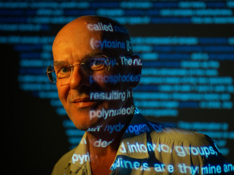
A new art exhibit at the Tompkins-McCaw Library for the Health Sciences at Virginia Commonwealth University features microscopic images of such things as a black widow spider's silk-spinning organ, mouse lung cells, muscle fibers within the inner ear and blood vessels from rat brain tissue.
All of the images in the exhibit, "Through the Looking Glass," were created by VCU students, faculty and staff. It is open to the public and will run from today until Dec. 31.
"When I was at O'Hare Airport last year, I saw an exhibit of microscopic images on display from the Argonne National Laboratory," said Teresa L. Knott, director of Tompkins-McCaw Library. "It occurred to me that we have microscopy facilities on both campuses along with students, faculty and staff who could create beautiful, scientifically interesting images."
A panel of judges from VCU's biomedical engineering and clinical laboratory sciences, led by Scott Henderson, Ph.D., director of VCU's Microscopy Facility, which is part of the Department of Anatomy and Neurobiology in the School of Medicine, chose the exhibit's 24 images from 40 submissions, and awarded three prizes on the basis of aesthetic appeal, technical skill and scientific significance.
"Exhibited images span disciplines from biomedical engineering to forensic science to pathology and range from scaffolding from electrospun fibers to a black widow spider spinnerets to a time lapse image of vagus neurons," Knott said. "The images selected for the exhibit are dynamic and fascinating to me."
To honor the prizewinners and showcase the images, a reception will be held today, from 3 p.m. to 4:30 p.m., at Tompkins-McCaw Library. It will be free and open to the public.
The winning submission, by Anders Hånell, a doctoral student in anatomy and neurobiology, is an image of a traumatic brain injury in a mouse.
"The axon in the center of the image, the long slender yellow structure going from the center to the bottom right corner, has a swelling near the center of the image," Hånell said. "At this point the swelling is only seen as a modest increase in the diameter, but they expand over time and eventually cause the axon to disconnect. Since the swelling and eventual disconnect occurs after the actual injury there's in theory it is possible to prevent it, but so far there are no treatments which can do this."
Ryan Clohessy, a student in biomedical engineering, created another prize-winning entry, depicting fibers of a biomaterial scaffold under a scanning electron microscope.
"The product has features at the nano and micro scale, and can be used to repair wounded tissue," he said.
Clohessy added that he feels honored that his work is featured in the show.
"I think combining science and artistic creativity has led to some amazing images and hope the beauty that can be found in science continues to be recognized and appreciated," he said.
Subscribe for free to the weekly VCU News email newsletter at http://newsletter.news.vcu.
Subscribe to VCU News
Subscribe to VCU News at newsletter.vcu.edu and receive a selection of stories, videos, photos, news clips and event listings in your inbox.